Wrist Fracture Surgery
The transcript below is YouTube's automatic one. Anatomy terms are the words the machine heard, so a few of them are wrong. The practice is proofreading them.
- Video ID:
rTpzfHbc3p4 - URL: https://www.youtube.com/watch?v=rTpzfHbc3p4
- Uploaded: 2014-11-26
- Duration: 668s
- Views at capture: 224104
- Captions: YouTube auto-generated (no manual track published)
Description (channel copy)
http://www.handandwristinstitute.com/wrist-fractures-surgeon/ - Dr. Knight looks at various fractures in the wrist and surgery to correct them. He specifically looks at the most common fractures: Distal Radius (Colles) and Scaphoid (Navicular) fractures. He also looks at more rare fractures of the Hamate and Capitate. He shows actual surgery footage of the top daredevil in the world, Red Bull motocross super star Robbie Maddison.
Displaced bones of the wrist almost always require surgical intervention, usually in the form of pins if the displacement is small enough to be put back manually, but often requiring plates and screws to hold the bone in place if the displacement is very great. These plates and screws are generally left in the hand, and do not impede function, but in the case that adhesions develop on the hardware, or if they somehow cause irritation or damage to the nearby tendons, they may be removed at a later date. Surgeries of this type, involving a plate, usually take about 45 minutes, and are done under general anesthetic. After this, a smaller surgery may be required to remove the pins, which is done at a later date.
Scaphoid fractures can be complicated, because the bone itself is not very well connected to the blood vessels of the hand, and so is delicate and susceptible to injury, and bone repair is difficult. Often, if there is enough vasculation in the bone, the scaphoid will heal itself without the need for surgery, but if it does not, then the bone can deteriorate, and must be either repaired removed. IF it is not removed, then a tension screw is used to hold the bone in place as it heals, and this can take five or six weeks.
In a hamate hook fracture, the best course of action is to remove the affected hook entirely, which allows the wrist to then move about freely without irritation.
Post surgery, splinting is the most important part of wrist recovery, and is necessary in all procedures to keep the wrist from moving and upsetting the delicate balance of bone placement for healing purposes.
Transcript
Let's take a look at wrist fracture surgery. Common cause of wrist fractures fall on an outstretched arm. Look at this snowboarder falling full weight impelling himself on the arm. So what are the symptoms? Let's take a look at this next image.
typically grabbing the wrist, significant pain when you break your wrist, swelling, bruising, deformity in severe cases, really severe cases, occasionally the bone may come through the skin. So again, all of these are are very worrisome when you have a lot of
pain for a wrist fracture after that type of injury. Let's look at the anatomy of the wrist. These are the carpal bones. There are eight bones that sit on top of the two forearm bones, the radius and the ulna. The most common fractured uh fractures in the wrist
involve the radius first and the scafoid second. These are the two most common common bones we're going to concentrate on. But first, let's look at the mechanism of injury for a distal radius fracture again or a scafoid fracture. A fall on an outstretched wrist. Forces
going up as you can see here through the palm shearing or pushing the wrist backwards. And as you can see here in this next slide, let's look at actual Collie's fracture, a distal radius fracture. Most common bone at the tip of the forearm bone is a radius fracture.
Now let's take a look at an actual displaced radius fracture. So fractures usually are two types. Either either they're normally aligned. If they're not, you got to get them normally aligned. In a kid, you can usually pop them back in place and they'll stay. In
an adult, usually it takes surgery to correct the problem. So, here's a displaced fracture. The ends are not lined up at all. And we're going to show you now the next image showing the bones realigned or what we call anatomic alignment. Again, in surgery, you're
knocked out for a about 30 minutes. We put these back in place and then frequently we'll actually cut open the skin and go in and put a plate and screws as you can see here on the front of the wrist. Okay. Now, let's take a look at an actual patient. This is a CAT
scan. Normally we get X-rays, but frequently the X-rays may show a a fracture, but it's normally aligned and we'll get still get a CAT scan because we'll see little cracks running up towards the wrist joint. And the wrist joint must be pristine. It must be put
back in place or is normally aligned as as perfect as possible because any inongruency in the wrist joint is like a pothole on a on the pavement here in Los Angeles where the wheels of the car go over that pavement and can wear down the joint. So, we want to get that as
perfect as possible. This patient, you can see the end of the radius. The arrow is on the palm side pointing towards the radius. And look, and these are the carpal bones out here. Look at these pieces. It's several pieces like an eggshell exploding. And these pieces to
have a good result need to be put back in place. So, let's take a look at this what we did to this patient. We went in put in a what we call a plate. That's along the or palm side of the forearm. We went in put this plate and screws putting everything perfectly back in
place and this patient had a very good result. Now let's take a look now at a at a uh typical Collie's fracture though where it's bent in a backwards position. The L shape the L on this is towards the dorsal side. This is the palm side here. Normally the bone should be tilted
slightly in a palmer direction this way. And in this case it's pushed backwards. So, we have to go in and push this back in place. Frequently, I'll go in and put a dorsal plate. You can put a plate, but the reason I like a dorsal plate in this patient is because we got a CAT scan
that showed a split into the joint. It's actually easy to fix when there's a fracture in the joint or ligament damage. If you go through the dorsal side, so you can see in this patient, the next image is going to show plate and screws on the back of the wrist, the
dorsal side of the wrist. And we actually put two little wires in because we couldn't get these the joint pieces back in perfect alignment without them. You can see this crack going into the joint and this helps keep that in place. Now the pins we have to take out about
five to six weeks later. A little minor surgery, but another trip to the operating room just to take those out. The plate and screws is designed to stay in. About 10% of patients may develop some tendon irritation or some adhesions to that and longterm may need to have
the plate removed. But this is a titanium lowprofile plate that's designed to stay in. So let's take a look at actually now the pen the pins have been removed. You can see here the plate is on the back side of the wrist and that restoration of the tilt of the
distal radius is now towards the palm. Okay. So let's take a look now at a scafo the scafoid which is the most common carpal bone that's fractured. You can see here fracture through this. And this is the scafoid is the most important bone of the carpal bones
because it bridges the row of bones here and the proximal row of bones here and links those two together. So in a and the if you look at the next image, the circulation to the scafoid is very tenuous. You have all these arteries around the wrist joint. But look over
here on the left hand side, you can see the arrow pointing to the scafoid and the fracture. But just beyond that fracture is the simp is the circulation to the scafoid. It's not much. And when you break the scafoid, this proximal portion or what we call the proximal
pole can die and just fall a apart like a piece of chalk. Uh or at least take quite a while to heal. The scafoid bone can take several months to heal and sometimes doesn't heal at all. It's one of the uh it's the longest healing bone in the body, per se. Now, let's take a
look actually at at the surgical procedure for this. When do we do surgery? Well, if it's just a hairline fracture, it's not separated, you can go with a cast. It can take at least six weeks like most fractures, but in this bone, as I said, could take several
months. But if it's displaced, surgery's recommended. Also, in an elite athlete who doesn't want to be in a cast for 3 to four months, they may elect us to go ahead and put a screw in this, which can speed up the recovery time to about five to six weeks. Let's look at the process
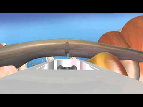
through a little um we first align the fracture, realign getting it back in its normal place. Then through a little uh puncture mark in the skin, we're going to put this guide wire in. And then over that guide wire, we're going to run this little screw in. It's a compression
screw that allows these that forces these two pieces together, which is optimal for fracture healing. Now, we're going to take the wire out. Now, we're going to see actually an X-ray image or an animated image of what this looks like. You can see this cool picture here
showing that screw in place compressing this. But let's look at an actual human being. This is a CAT scan showing the arrows pointing on the palm side. This is the dorsal or back side of the of the we're going to look at it from the side view. And then you can see a crack going
through the waist or middle portion of the scafoid. How did we fix this? We put a screw in. Let's take a look here. This is the actual patient. This is this patient about six weeks later. Everything's healed. You can see the screw in. No more crack is visible. And
this patient's getting back to normal activities very quickly. So, let's take a look now at a at a at another carpal fracture, not near as common as as these other two bones that we just talked about. It's called a hammade fracture. The hammade is is a bone that has a
little uh hook or protuberance that's coming off the palm side here. And when you fall on an outstretched wrist, striking the palm on the ground or using racket or club sport such as baseball or something where or golf where you drive the club into that bone, it can shear it
off. So frequently it'll take an MRI to see this and you can see actually the base of this is the hammate bone right here. You can see this gap is is where the bone is not healing and the tip of the bone here. All we do for this is we go in and simply remove the tip of the
bone and start you know recovery right away. Not a long period of immobilization like most fractures because we're simply removing the tip of the bone. It doesn't have a lot of function. So we get it out of there because if it stays in there and stays
painful and symptomatic actually can rupture the tend the tendons to the little and ring finger and cause a lot more uh grief in the long haul. And next uh fracture we're going to look at is called a capotite fracture. Very uncommon fracture except in very violent
injuries. The f the next image I'm going to this image I'm showing you right here is Robbie Madison, one of the the modern-day evil conval Red Bull athlete who came to me several years ago after he had fallen about 50 feet on uh the track out of doing a stunt came down
impelled his wrist right into the ground and after several misdiagnosis we got a CAT scan and uh and we could see actually you can see here the fracture the capitates right here. This is the tip of the capitate with the metacarpal bones out here to the uh third middle
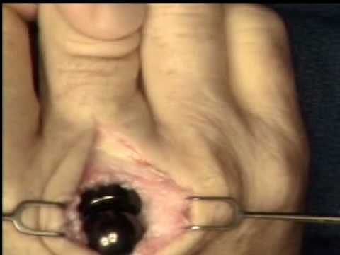
finger. And he sheared this off. Let's take a look actually at his surgery. We go in right here. You can see we've put everything back in place. We've put these pins in right here and here. I'm showing the smooth alignment of the capitate. Everything's back in place.
We're going to take out this retractor. And we're basically just going to snip off the uh the wires right here underneath the skin. As you can see here, we're going to take a little biter and we're actually going to clip that off. Again, he had three pins put in.
We're going to clip these off. We're going to leave them under the skin for about 5 to 6 weeks until it's healed. Then a simple procedure to go back. As you can see, the wire's gone. Now, let's take a look at actually at Robbie's postsurgery X-rays. Right after surgery,
we get uh we actually do this with real time what called fluoroscopic imaging. And you can see here there three wires. Here's the capitate perfectly aligned. But let's take a look after the pins came out at a CAT scan of Robbie's wrist. You can see here perfect
alignment of the capitate bone. This guy is amazing athlete. Five weeks out, he had the pins out and I said to take his time recovering, already had a graphite brace on and was back to having other body injuries by the by the daredevil stunts he performs. Now, let's take a
look at u actually after these surgeries. Normally we place you in a splint postoperatively for a brief period of time and then after the healing we or even in a non-displaced fracture that we don't do surgery on. We usually go to a fiberglass cast. We
change it every two weeks with with new X-rays just to make sure everything's healing. Okay. You can see here it's a thumb spica cast that actually comes out on the thumb. This is typical for a scafoid fracture because it's been shown if you immobilize a thumb it leads to a
better healing rate or quicker healing period. Sometimes the this cast will extend above the elbow in certain fractures like a bad distal radius or colleies fracture or an escafoid fracture. Some may elect to put the cast all the way above above the elbow but
usually in most uh most patients it's about a six-w weekek recovery. For more on this and many other conditions, please take a look at our website.