Endoscopic Carpal Tunnel Release (ECTR)
The transcript below is YouTube's automatic one. Anatomy terms are the words the machine heard, so a few of them are wrong. The practice is proofreading them.
- Video ID:
rp9HwTGroXY - URL: https://www.youtube.com/watch?v=rp9HwTGroXY
- Uploaded: 2010-07-04
- Duration: 628s
- Views at capture: 53667
- Captions: YouTube auto-generated (no manual track published)
Description (channel copy)
http://www.handandwristinstitute.com/carpal-tunnel-release-surgery-los-angeles/ - Video of stitchless endoscopic carpal tunnel release. This is the most advanced carpal tunnel release video.
Transcript
Over 2 and a2 million people a year are inflicted with the diagnosis of carpal tunnel syndrome. To understand this diagnosis in better detail, it's important to understand a little of the anatomy in the wrist. The carpal tunnel is a tunnel deep in the wrist here
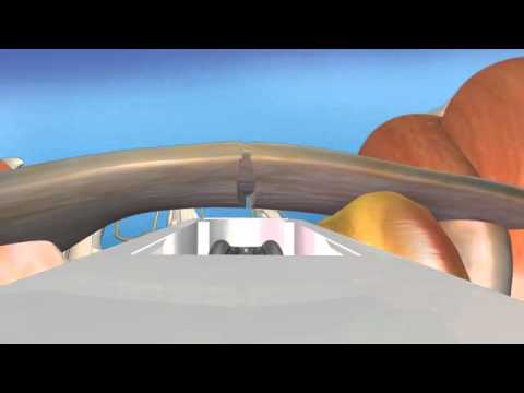
that's made up of a the t the floor of the tunnel is made up of the back of the bones of the wrist. Across that tunnel is a ligament called the transverse carpal ligament which we'll talk about when we talk about the surgery for this condition. But with technology today,
there's increased use of the fingers flexing through that tunnel that leads to a development of inflammation on the tendons. Within that tight, unyielding space, the inflammation puts pressure on the median nerve, which is a nerve that goes through the tunnel out to the
fingers of the hand. Within from repetitive use with daily activities, with uh increased computer use, with Blackberries, etc., Our technology has made this a more common occurrence. To discuss a little of the diagnosis of carpal tunnel syndrome, the
subjective complaints or when a patient comes into my office, one of the first things they talk about is that they have numbness and tingling, more commonly awakening them at night. Commonly, it occurs because the nerve goes to these four digits. It's mainly the four these
four digits excluding the little finger. They also with more pronounced symptoms may get actually loss of feeling in their hand and actual motor loss of the of the muscles that allow the thumb to oppose to the digits resulting in clumsiness with buttoning close using a
pen for writing etc. Under examination the patient we tap over the nerve at the tunnel the median nerve and if that causes tingling within the fingers that's suggestive of the diagnosis. We also ask the patients to flex their wrists leading to
increased pressure within the carpal tunnel which is a called a a failins test. So that reproduces what happens at night when you're sleeping. You tend to sleep in a fetal position the wrist drawn up leading which causes increased pressure within the carpal tunnel. Also
we look at the we do a full sensory and a full sensory and motor exam to see if there's loss of feeling and any loss of strength within the thumb muscles. So once we have a proper diagnosis of carpal tunnel syndrome now it's important to talk about treatment.
Treatment in most cases with conservative treatment the symptoms will go away. In severe cases though surgery may be necessary. For the conservative treatment wearing a wrist brace at night when you're sleeping to prevent flexing flexion of the wrist is important.
That's the number one treatment. also resting the wrist and the fingers ergonomically looking at the workstation at work if they're a typist or if or adjusting the string tension if they're a guitar player etc. Uh anti-inflammatory medication. In some
cases, a cortisone shot into the carpal tunnel may also reduce swelling. In more severe cases though, surgery may be necessary. Now, with the advent of endoscopic carpal tunnel release, surgery is highly effective, but with minimal scarring and a very quick return
to work, we do a 10-minute outpatient surgical procedure that involves a 1 and a half centimeter incision across the crease of the wrist. And the pain and the recovery, the pain is lessened significantly compared to the old-fashioned open procedure, which
involved a very lengthy scar across the palm. Now, I have patients returning to full work uh within a month after surgery. So, it's highly effective and much less pain than the than the standard open procedure. Now, we'll start with a skin
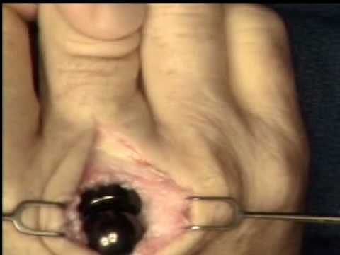
incision.
I'm going complete the cut
proximal. And now I have a U-shaped distalbased flap that we're going to now place a small skin hook on that will essentially be my guide into the carpal tunnel. Force, please. Right
here. Now I'm going to open approximately 2 cm of the anrachial fascia at the lower midline of the distal forearm proximal. That'll help facilitate the scope placement into the carpal tunnel.
Now we'll take the spatula and I'm going to run this into the carpal tunnel and I'm going to scrape the under surface of the transverse carpal ligament just to make sure there's no tenosovium or lining of the tendons
that's adherent to the under surface of where I'm going to be dividing the transverse carpal ligament.
So now we're going in into the carpal tunnel. You can see the transverse carpal ligament
fibers. Right
there you see I'm pushing beyond the transverse carpal ligament, beyond the carpal tunnel.
Now I'm going to engage the blade. You see right there? And I'm going to cut the not go all the way the very end. I'm going to cut the distal fibers here partially. See the partial cut there. Now this two edges you can see
are still intact there. So you got to if you see those within the viewing surface of the or within the video monitor and you haven't and it's a V-shaped configuration there. You haven't completely transsected it. Little fat on the There we go.
We're going to complete the transsection proximal. We're going to go all the way but not to the very edge proximal. We'll come back and do that with our scissors. Pistal. Now we we're going to run the radial column of the transverse carpal ligament
all the way proximal showing complete division except what we're going to complete proximal with our scissors on the way out and then there's the owner column. Again you don't see the two edges within the view of the scope at
any point. All right. So that's open all the way down and we're going to inspect the media nerve. Now you can through this field look at the nerve and make sure there's no masses within the carpal
tunnel. That's the nerve there and it's intact after the procedure which is important obviously. Okay, come on out. Now we're going to let just apply some uh pressure to the incision and we're going to let the tourniquet down. Now
tourniquet down please. It's usually with direct pressure for about 5 minutes is all that's needed to stop any oozing before closing the wound.
You can see the fingers pinking up now, all of them, and the palm. And I'll just liberally apply some Dermabine skin
glue. Dressing, please.
I'm just going to put a little little sponge with a Teoderm dressing over it so they can start showering right away.
And now they can apply ice post-operatively along with elevation to minimize swelling and can start range of motion of the hand and wrist within the first 24 hours. This procedure as you can see involved a very small incision through a minimally invasive approach as
you can see from her left wrist done 3 months ago. With this latest technology we are now able to bring life back to her hands.