Wrist Ligament Surgery
The transcript below is YouTube's automatic one. Anatomy terms are the words the machine heard, so a few of them are wrong. The practice is proofreading them.
- Video ID:
Pd8w09Q_gtM - URL: https://www.youtube.com/watch?v=Pd8w09Q_gtM
- Uploaded: 2014-11-01
- Duration: 308s
- Views at capture: 72625
- Captions: YouTube auto-generated (no manual track published)
Description (channel copy)
http://www.handandwristinstitute.com/cartilage-tears-wrist-doctor/ - As the hand and wrist are extremely delicate parts of the body, the ligaments that ensure the bones stay in the right position are very important for movement. Should these ligaments become disrupted or moved out of place, pain, and function loss can occur, as well as the premature development of arthritis. Most likely, these injuries occur when the patient falls on the outstretched wrist, which is often seen in athletic injuries. Because of the non-bony nature of ligaments, it is easy to miss tears to them on a standard X-ray, which is why it is important for patients suffering from them to see a specialist for more effective diagnosis and treatment so that the injury is not misdiagnosed.
Of the ligaments in the wrist, it is most common to see injury to the scapholunate ligament, which connects the scaphoid and the lunate, and the lunotriqutral ligament, which serves the same purpose for the lunate and triquetrum bones.
The aforementioned unreliability of traditional x-rays means that a more indicative test, the MR-Arthrogram, is generally necessary to determine injury to these tendons. The radiocarpal joint in injected with dye, and if the dye flows from there to the midcarpal joint, then a torn ligament is very likely. Even though an image like this will indicate injury in about 70% of cases, even a normal MRA does not necessarily ensure that the ligaments are not torn.
If the tear to the ligaments are only partial, then a four to six week regimen of splinting may be effective in allowing sufficient immobilization for healing. Should this prove ineffective, and if the injury is caught in time, then there is a three-month window in which an arthroscopic procedure may allow the injury to be corrected, and this procedure is found to be effective in 80% of cases.
Often, however, the damage to the ligaments may be more substantial, such that they cannot be reattached to the bone. If this is the case, a small vacuum-like tool is used to clean up the torn edges and scar tissue on the ligament, after which time tiny metal pins are installed in the wrist to hold the bones together and allow the ligaments to rebuild themselves and develop some scar tissue.
An eight-week period of immobilization is necessary after this type of procedure, facilitated with a brace. After eight weeks, surgery is undergone to remove the pins, and then three months of therapy are necessary to return use of the hand of full function. If diagnosed in a reasonable amount of time, this minimally invasive procedure should prove effective in reducing symptoms and returning to full function.
Transcript
let's talk about wrist ligament surgery the wrist ligaments are are small ligaments that hold the eight bones in the wrist together and they provide important tethering of these bones during wrist movement when just one ligament becomes disrupted this Alters
the mechanics of the wrist which can cause significant pain and significant Li limitation of function leading to premature arthritis over the long term this occurs most commonly from a fall and an outstretched wrist look at this snowboarder going down all his body
weight under the hard surface with that extended wrist frequently you go into the emergency room these patients get normal x-rays which is usually the case and this does not rule out a soft tissue or significant ligament disruption in many cases so it's frequently missed the
diagnosis is missed and after several months the patient may just present to our office when frequently it's too late now's look at the anatomy of the wrist this shows the carpal bones right here the forearm bone the IUS the Onna over to the left hand side you can see the
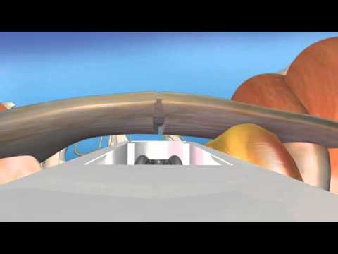
scaphoid bone here the sca this blue ligament uh called the scaal lunate ligament between the sca lunate and the scaphoid and over between the lunate and the triquetrum is a luno triquetral ligament these are one millimeter thick ligaments that tether these three bones
together very important structures but the most frequently uh Tor tears of ligaments in the wrist let's look at an MRI now this is called an MR orogram and it's the gold standard uh for diagnosing ligament tears in the wrist before performing invasive
orthoscopic surgery this shows the fluid this white fluid that's injected into the radiocarpal joint which is the arthrogram component of the test this flu this fluid should stay right along this joint it should not enter between these bones as you see it doing here and
you can see this arrow is pointing to A disruption of this black line which is the scaal lunate ligament now initially with a ligament tear on the wrist conservative treatment would be about four to 6 weeks in a cast or brace and sometimes that will heal but after that
period there's about a 3month golden period where we aggressively treat these with minimally invasive arthoscopic surgery to go in and try to get these ligaments to heal let's look at an actual normal scaal lunate ligament this is a lunate here Co this bone is covered
by normal cartilage and then over on the scoid the same and then there's this fluffy ligament where the arrow is pointing called the scapol lunate ligament that can be disrupted so let's look at an actual disruption here this is through again looking at the monitor
we're seeing in surgery through a 2mm cut we're putting in this tube that broadcasts through a camera to this monitor we're seeing right here this image through a second 2mm cut on the back of the wrist we're putting in this small probe that looks magnified looks
large here but it's really a very fine probe that is actually coming in and hooking behind the scao of the scao lunate ligament pulling it away from the scaphoid it's a pretty substantial tear now usually these ligaments are torn down the middle in which case we um they
don't have the ability to be repaired sometimes though they're pulled off the edge of the scaphoid or the lunate where you can actually go put an anchor in place and tie these down but in this case minimally invasive surgery ligament is uh as we said is frequently torn down
the middle so what we do is we go in and put three stainless steel pins between the carpal bones where the ligaments torn the scoid the lunate three stainless steel pins are placed in these are cut off right underneath the skin but this shows again normal alignment of
the scoid and the lunate now the next most uh common ligament to be torn is the luno triquetral ligament you can see the luno triquetral ligament is this depression here between the triquetrum and the lunate down in the bottom leftand corner but it's in this area as
you can see this can be torn well let's look at a tear this is a little grainy but you can see here the triquetrum and the lunate where the L is and you can see this Gap right here and so what we want to is we want to put these pins in that will be in for a period of time
that will allow the these ligaments and the bones to adhere together to Res restore stability to these uh hypermobile uh bones so let's look at actually the the again pins coming in from the opposite side of the wrist with this less commonly torn luno triquetral
ligament we put three pins here that rigidly hold these bones together now these pins will stay in for about eight weeks and then through another little five minute outpatient procedure uh we take the pens out and we start rehab it's a very carefully orchestrated
rehab protocol that we follow for about three months before return to normal function the success rate is 80% when it's in that three-month golden period each month that goes beyond from that uh becomes less successful but overall the success rate is much greater than the uh
much larger more invasive reconstructive procedures that are done for more chronic injuries for more on this condition and many conditions please visit the Hanah rist Institute website