Mucoid Cyst Surgery
The transcript below is YouTube's automatic one. Anatomy terms are the words the machine heard, so a few of them are wrong. The practice is proofreading them.
- Video ID:
VUsOfmV_uTw - URL: https://www.youtube.com/watch?v=VUsOfmV_uTw
- Uploaded: 2014-11-15
- Duration: 231s
- Views at capture: 134146
- Captions: YouTube auto-generated (no manual track published)
Description (channel copy)
http://www.handandwristinstitute.com/digital-mucoid-cysts/ - When ganglion cysts occur at the tip of the finger or in the nail bed, they are referred to as mucoid cysts. Unless they are close enough to the nail to cause deformity, they generally appear as a small, simple bump on the finger. Appearance of these cysts is generally associated with occurrences of arthritis in finger joints, but most often it is the cyst that causes pain, and not the underlying arthritis. Cysts most often occur on joints where bone spurs appear, which lead to the formation of the cysts in the first place.
Arthritis and bone spurs are best diagnosed using x-rays, but it is also possible that MRI imaging may be necessary to get a better look at the cyst itself.
People often treat cysts by aspirating them, but this very seldom leads to resolution of symptoms, and the cyst can often grow back in a very short time, so that surgery is the preferred method of resolution. In almost all cases, surgical treatment involving the complete physical removal of the cyst and related parts, as well as bone spurs, will lead to the complete resolution of symptoms. Extensor tendons are delicate and must be carefully avoided during surgery, otherwise mobility and use of the hand may be affected. It is also important that the surgery be undertaken prior to any outward eruption of the cyst through the skin to avoid infection.
Once surgery is completed, therapy should be initiated as soon as possible. After about ten days, the external sutures holding the wound together will be removed, and the overall recovery time should be about four weeks. Recurrences of this type of cysts, post-surgically, occure in less less than 5% of cases.
Transcript
let's take a look at mucoid cyst surgery a mucoid cyst is a small gangan cyst that arises from The Joint out called the distal inter flangel joint or dip joint which is right out here near the nailbed let's take a look you can see here just a bump right over the joint
and over here you can on the left it looks just a simple bump right over the over the joint whereas on the right side there's actually a nail Groove happening and why is that because the cyst although it communicates with the with the joint it actually has tracked and is
formed out here over the the germinal nail Matrix which puts pressure on the area that forms the nail leading to a linear or depressed groove of the nail deformity which actually will resolve um after surgery in many cases so let's take a look now at actually what's
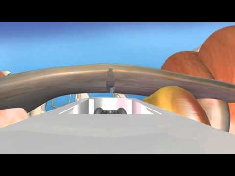
happening Under the Skin this shows a nice schematic of the distal interial joint between the distal failinks at the tip and the middle failinks right here and the out of the joint capsule from underlying arthritis pops of cyst almost like a little mushroom type uh structure
filled with this jelly and it can fluctuate ins size as the fluid can drain back and forth between the joint and the cyst so also associated with this as I said was is degenerative arthritis but let's take a look at the bone spurs that can form right
underneath the cyst and these are important as we'll talk about in a little bit to take these out to lower the recurrence rate when surgery happens okay so let's look at now at this next image which is actually an X-Ray when when you come in when you have a bump on
your finger well you'll think it's just a a little mass and why get an x-ray well the reason we want to get an x-ray is to is to see if there's an underlying bone spur from that underlying arthritis this patient has mild arthritis but in the circle area where the arrow is
pointing you see a little [ __ ] of bone or calcification and that's a bone spur so when we go in to remove that Cy it's important to take that out and some patients an MRI will be obtained as you can see from this next image the mucoid cyst labeled Arrow is pointing to this
white structure right over the over the distal interial joint or dip joint and this is filled with fluid show it shows up on this MRI is a white structure again just to further um diagnose what's going on in there and distinguish this Mass from others so after this after the
MRI is taken usually this is a surgical cure in this patient you can see a large bump right over the distal inter fenial joint There's real no there's no nail plate abnormality so in in a lot of patients they will have seen a dermatologist or other doctors will have
gotten multiple cortisone injections or sucking the fluid out of the out of the the cyst that can provide temporary relief but usually that's not Curative unless you go in there remove the cyst and take out the Spurs let's take a look we're going to cut down through the skin
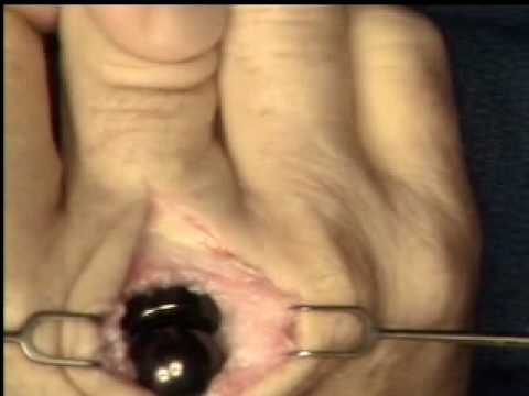
you can see this magnified view great picture of this large cyst that you just saw before the skin was cut open but now you can see multi-lobulated all these different like balls together all filled with this gelatinous fluid fluid over on the top side you see this white
structure running along here that's the extensor tendon that's the tendon that straightens the finger so we be very careful to go right off the edge of the tendon take out the cyst and then there's going to be a little bone spur as there was in this patient we just
nibble that off a little biter and then we sew the skin together so those stitches are going to be in after this little dressing the stitches will be in for about 10 days you come back to the office after the initial wound check 10 days later to get those stitches out
you'll start ranging motion right away after surgery to prevent stiffness therapy in some cases overall This is highly successful surgery about a 95% success rate and there's about a less than a 5% chance of recurrence for more on this condition
and many other conditions please check out our website